Repository
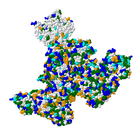
7TEQ
This image shows a Fab5 antibody fragment (PDB 7TEQ) from Mus musculus (mouse), determined by X-ray crystallography at 2.30 Å resolution.
Image rendered by: Lara Hernández Paredes

5ERM
The crystal structure of the cyclization domain of Phomopsis amygdali fusicoccadiene synthase is presented in PDB 5ERM. This structure is complexed with magnesium ions and pamidronate, highlighting the enzyme's active site configuration.
Image rendered by: Emilio Ruiz Molero

7AYS
The structure of bovine trypsin (PDB 7AYS) was determined using single femtosecond snapshots at room temperature. This enzyme is a serine protease involved in protein digestion.
Image rendered by: África Paredes Martínez del Peral
Loading...